Educational Review—Improving Surgical Outcomes Cost-Effectively

STUDY #1: Intraoperative CT Improves CI Surgical Outcomes
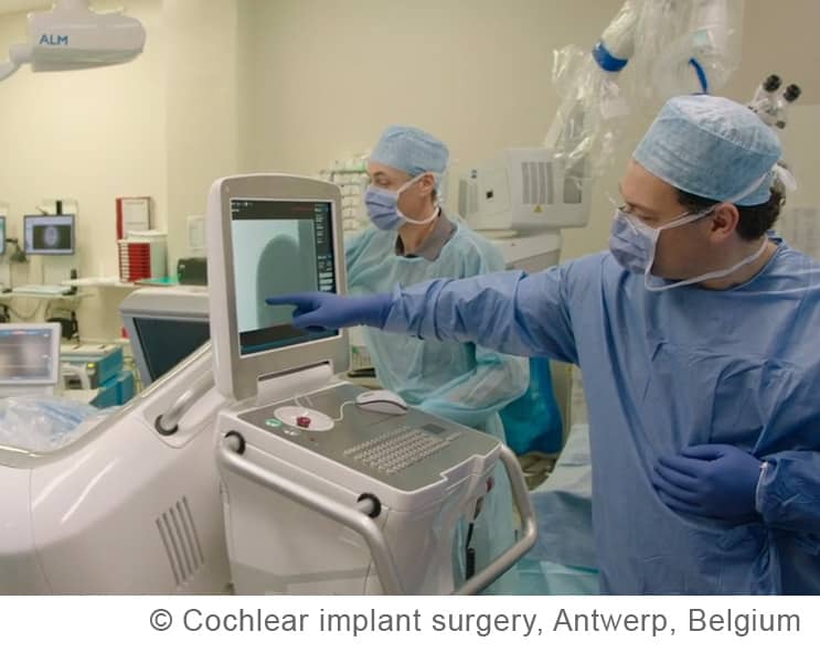
In a recent published study by Vanderbilt University entitled, “Use of intraoperative CT scanning for quality control assessment of cochlear implant electrode array placement,” clinical research using the truly mobile xCAT XL shows how intraoperative CT scanning in cochlear implant surgeries has improved electrode array placement outcomes for patients.

“3D imaging, a.k.a. CT scanning, affords a new level of quality assessment by not only identifying misplacement and/or tip fold-over but also providing detailed information about intracochlear position including scalar location and distance from the modiolus.”
Robert F. Labadie, M.D., Ph.D,
Department of Otolaryngology-Head and Neck Surgery, Vanderbilt University, Nashville, TN
Better Cochlear Implant Positioning from Superior CT Visualization

As a first step, we have shown preliminary data that intraoperative CT scanning has resulted, over time, in surgeons becoming better at achieving perimodiolar positioning which is known to be associated with better audiological outcomes.”


