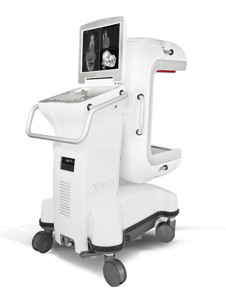
Xoran’s VetCAT IQ 3D CT scans identify foreign body
A two-year-old female canine presented with a history of intermittent discharge from the right eye. Visual® examination identified limited mobility of the third eyelid, but no evidence of a foreign body. Medical treatment was first considered, but because the VetCAT IQ was readily available, a high-resolution soft tissue CT acquisition was ordered.
The CT image results pointed to the presence of a foreign body. An orbital surgery was performed and a 2 cm long wood fragment was retrieved from the ventral orbit.

VetCAT imaging shows the extent of bone loss and nasal changes caused by the oronasal fistula.

Patient’s eye with discharge during this initial examination

Intraoperative image of the 2 cm piece of wood embedded within the conjunctiva

The VetCAT IQ provided immediate 3D CT images
of the boney anatomy and tissue, showing the
area of concern in the right orbit.
Without VetCAT IQ, the irritant would have been missed. The ability to bring this high-resolution mobile CT to the treatment table during the initial evaluation is very significant.
This technology allows for a ground-breaking intraoperative imaging capability without
moving the patient from the operating room.
Total time to obtain the CT, including positioning of VetCAT IQ is about 4 minutes.

Courtesy of Sinisa Grozdanic DVM, PhD, and Diplomate — American College of Veterinary Ophthalmology


