VetCAT enhanced 2D & 3D imaging provides clear visualization of draining tracts within the mandible
VetCAT 3D scans helped guide optimal treatment planning

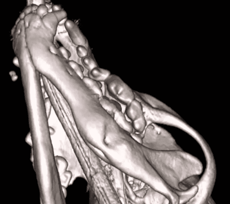
Piper, a two-year-old male Yorkie, presented for a routine dental cleaning. The referring veterinarian was surprised to find multiple periapical lesions. A 3D reconstructed image from the cone beam reveals draining tracts from the mandible. Dr. Bannon was able to successfully extract the tooth without fracturing the mandible with the help of VetCAT CT imaging.

VetCAT imaging shows the extent of bone loss and nasal changes caused by the oronasal fistula.

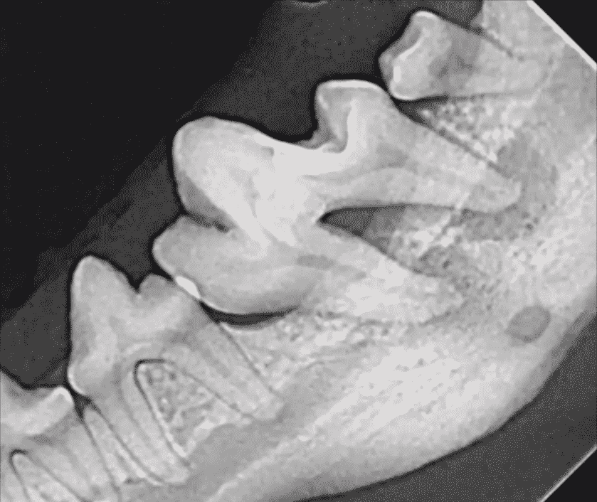
Visible periapical lesions with dilacerated roots are seen here in the 2D radiograph.

With the cone beam’s 3D reconstructed image, the periapical lesions are clearly visible along with two draining tracts—one out of the ventral cortex and one out of the buccal bone plate.
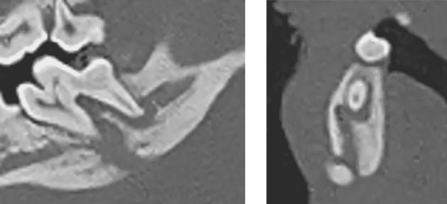
Visibility of the draining tracts in the cross-sectional views show communication of the draining tracts between the buccal and lingual plates.

“The cone beam image gives much more visibility, which really helps the owners and veterinarian make a better plan.”
– Dr. Kris Bannon
Xoran is passionate about helping animals and supporting veterinarians


