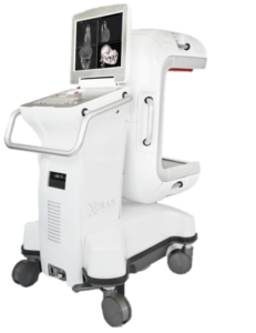
Xoran’s VetCAT IQ 3D CT scans can prevent unnecessary surgery
A five-year-old male canine presented with a history of facial swelling. The patient developed swelling in December 2021 in the area of a discolored tooth. After his regular veterinarian extracted the tooth, the swelling did not improve, even with antibiotics and steroids. A fine needle aspirate was not diagnostic.

VetCAT imaging shows the extent of bone loss and nasal changes caused by the oronasal fistula.
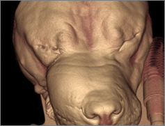
3D enhanced view showing facial swelling

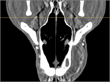
Soft tissue CT scan with contrast shows the extent of the swelling and ample blood supply


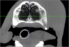
Bone window CT scan shows abnormal changes and edema in the turbinates under the swelling

A small incisional biopsy was taken while the patient was under anesthesia, and two scans were performed: a bone scan, followed by a soft tissue scan with contrast.
The incisional biopsy came back a few days later as a maxillary fibrosarcoma. When presented with a very extensive and invasive surgery with a high probability of recurrence, the owner elected only palliative care.
“It is a good example where having excellent imaging allowed us to make an informed decision not to treat. Prior to having CBCT we might have been tempted to go after the tumor, only to find out from the histopath after the fact that we didn’t achieve adequate margins. Radiographs weren’t terribly helpful in this case.”
– Dr. Hutt
Review courtesy of: Jason Hutt, DVM Animal Dental Centers


