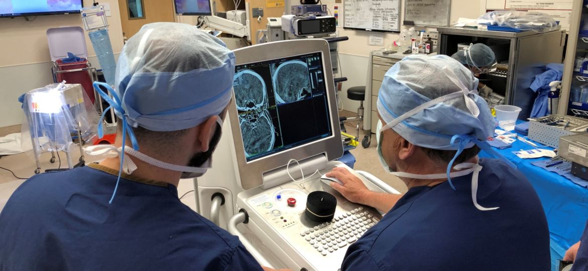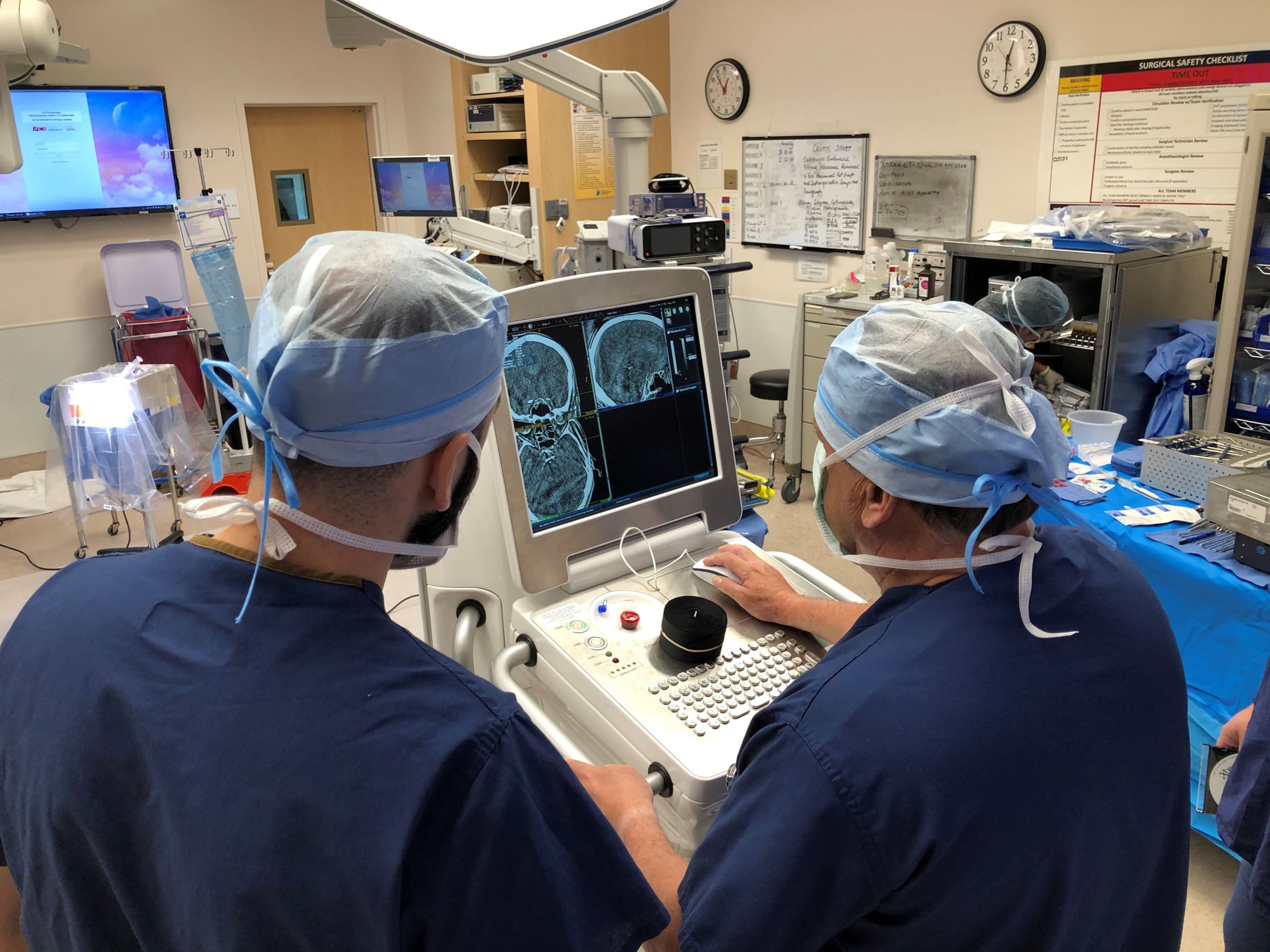

Ann Arbor, MI, September 23, 2021 – Xoran Technologies announced that the company is conducting a clinical study to document a series of cases using Xoran‘s xCAT IQ mobile CT scanner. In partnership with Pacific Neuroscience Institute (PNI), the intent of the study is to demonstrate that the selected techniques and resulting image quality are adequate for the clinical assessment of common neurosurgical conditions.
The study includes patients undergoing complex cranial neurosurgical procedures which can greatly benefit from low dose intraoperative imaging—allowing surgeons to view anatomy during surgery and confirming surgical completeness. These procedures can include endonasal pituitary and skull base tumor resection, and open craniotomy treatment of tumors such as meningioma, glioblastoma, or acoustic neuroma, ventricular shunts, and/or neurosurgical procedures for hydrocephalus, deep brain stimulation electrodes, subdural hematoma evacuation, or ventricular endoscopy.
The study is designed to examine benefits associated with easily obtained CT imaging of the head at the point of care, instead of transporting the critical patient to a conventional scanner. Other points of interest include the ability to obtain intra-operative imaging prior to procedure completion, the potential for reduced radiation exposure, ease of use, and ease of access to imaging.
Principal investigators of the study include Chester Griffiths MD, FACS, Professor of Surgery, and Garni Barkhoudarian MD, FAANS, Associate Professor of Neuroscience / Neurosurgery. Co-investigators are Daniel Kelly MD, John Rhee MD, Jean-Philippe Langevi MD, and Walavan Sivakumar MD.
“We are very pleased to support doctors Griffiths and Barkhoudarian, PNI and their colleagues—to see the xCAT IQ used intraoperatively, advancing the field of medical imaging in support of these complex neurosurgical cases,” said Laura Dennis, Xoran’s Vice-President of Sales and Marketing. “Xoran is committed to meeting the needs of today’s surgeons, helping them provide real-time low dose imaging, in the OR and the critical care unit.”
xCAT IQ is a mobile CT scanner that can be positioned in the operating room to acquire cranial images at the time of surgery. This device, which resembles a large cart, is relatively compact, can be pushed into position by a single operator. Once in place, acquisition of the CT scan takes about 40 seconds and image reconstruction takes a couple of minutes.
With a two-decade track record of innovation in medical CBCT imaging, Xoran understands the CT needs and practical considerations in both the OR and office settings.
###
ABOUT DR. GRIFFITHS
Chester F. Griffiths, MD, FACS, is board certified in Otolaryngology, Head and Neck Surgery and Facial Plastic and Reconstructive Surgery. He has an extensive 30-year experience in endoscopic endonasal sinus surgery for skull base tumors and pituitary tumors, sinonasal cancers including mucosal melanomas, and in the treatment of facial and nasal trauma, cosmetic deformities, sinus infections, and disorders of smell and taste. His practice also includes treatment of sleep apnea, snoring, difficulty breathing, disorders of the larynx, thyroid tumors and other head and neck cancers with an emphasis on viral HPV related cancers.
More at https://www.pacificneuroscienceinstitute.org/people/chester-griffiths/
ABOUT XORAN TECHNOLOGIES
Since 2001, Xoran is the pioneer and medical market leader in low-dose radiation, cone beam CT systems specifically designed for the patient’s point-of-care. Providers around the world rely on our industry-leading MiniCAT™, xCAT™, VetCAT and vTRON systems to diagnose and treat patients. Xoran is based in Ann Arbor, Michigan.
For more information visit www.xorantech.com/xCAT
Pacific Neuroscience Institute www.PacificNeuro.org
© 2021 Xoran Technologies, LLC
Media Contact
Aramide Boatswain
+1.734-709-0464
info@xorantech.com


