Make the Transition from 2D to 3D… Improve Diagnostic Capability and Outcomes
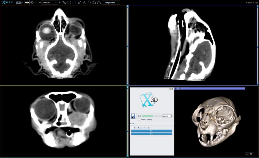
CASE OF THE MONTH: Use of VetCAT IQ for soft tissue diagnosis of orbital tumor

View a feline primary orbital tumor with real-time 3D soft tissue imaging.
Show clients the extent of 3D image intraoperatively to view
the disease and to determine the best treatment.
Conversations to have with clients—
Orbital tumors are best seen on 3D imaging. The extent and type of tumor can typically be seen more clearly.


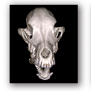
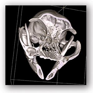
Dog anatomy can be rotated and cropped in 3D. The floor of the orbit, zygomatic arch and mandibular ascending branch are observed.
Pathology and possible communication with other compartments such as dental apices or the oral cavity. The patient above shows a well-defined cyst in the right maxilla, which is clearly not invading the orbit.
Sources: xorantech.com/veterinary-references
ABOUT DR. GROZDANIC
Dr. Grozdanic is a Diplomate of the American College of Veterinary Ophthalmology with expertise in glaucoma, retinal diseases, and cancer-induced ocular complications. Dr. Grozdanic published close to forty scientific articles, one textbook and gave almost two hundred talks to veterinarians all over the world. In collaboration with Xoran, his current research interests include intraoperative CT imaging and stereotaxic orbital surgery for dogs and cats.


