VetCAT 3D Imaging Creates 3d Printed Mandible for Mandibular Reconstruction Planning
VetCAT 3D scans help guide surgical planning
Wilby is a ten-year-old male Golden Retriever who presented with severe mandibular drift from a previous mandibulectomy from canine acanthomatatous ameloblastoma (CAA). With the help of VetCAT 3D reconstruction, Dr. Goldschmidt was able to recreate a 3D printed mirror image of the mandible to help surgically plan Wilby’s mandibular reconstruction.

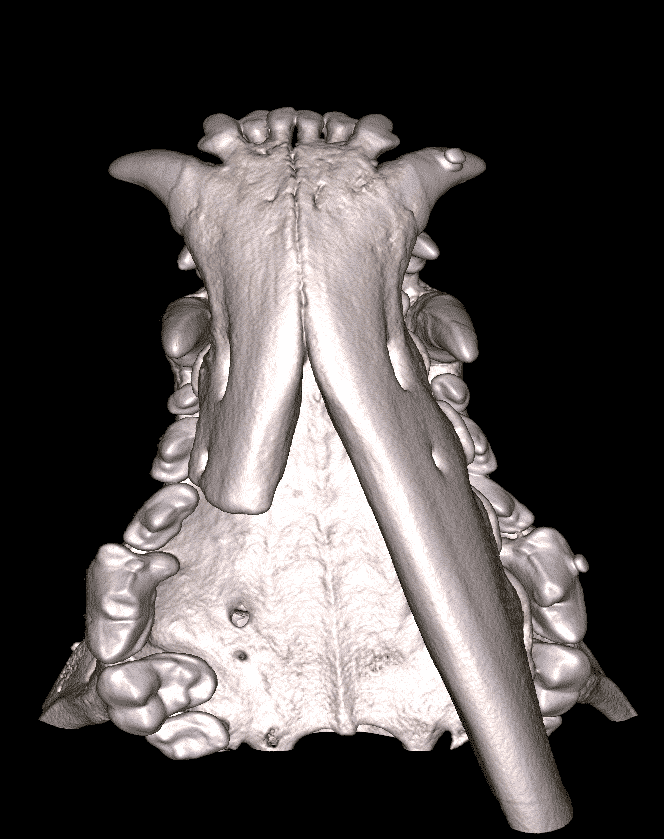
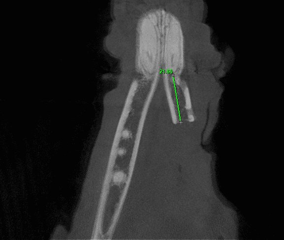
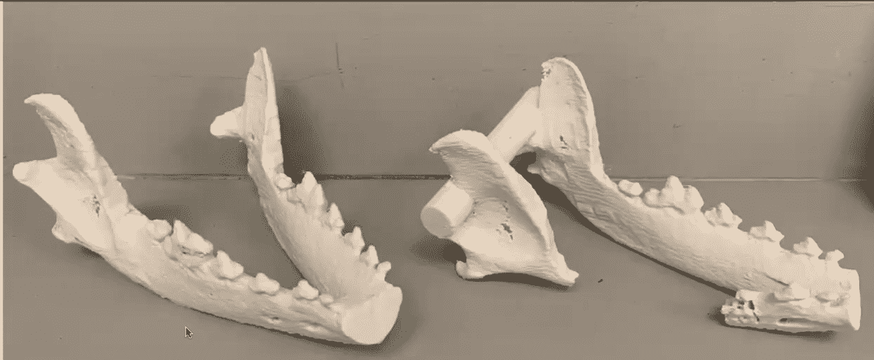
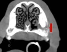
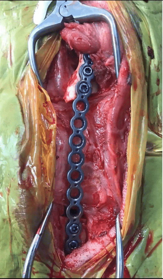
Wilby had previously undergone a mandibulectomy for CAA and has since suffered from continuous soft tissue trauma and discomfort. The scans above show the VetCAT’s 3D reconstruction of the defect, and measurement from the apex of the canine tooth in the remaining mandibular bone using VetCAT software. A 3D printed model was created from the cone beam scan data, they were able to print a mirrored image of the non-defect side to create a model of what the mandible should look like. This helped the planning and fitting for the type and size plate they would use to reconstruct Wilby’s mandible. The image on the right shows the plate installed intra-operatively.
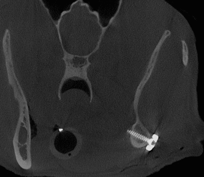
An immediate post-operative cone beam scan was performed after surgery. Imaging obtained helped Dr. Goldschmidt and the team feel confident that the screws were in alignment and the apex of the tooth had not been disrupted and no emergency root canal was needed. Dr. Goldschmidt was able to plan Wilby’s entire surgery based off the VetCAT’s cone beam imaging.
Xoran is passionate about helping animals and supporting veterinarians


