Dental Injury
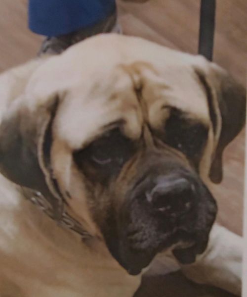
Jericho is a 5 year old Mastiff with a LOT of character.

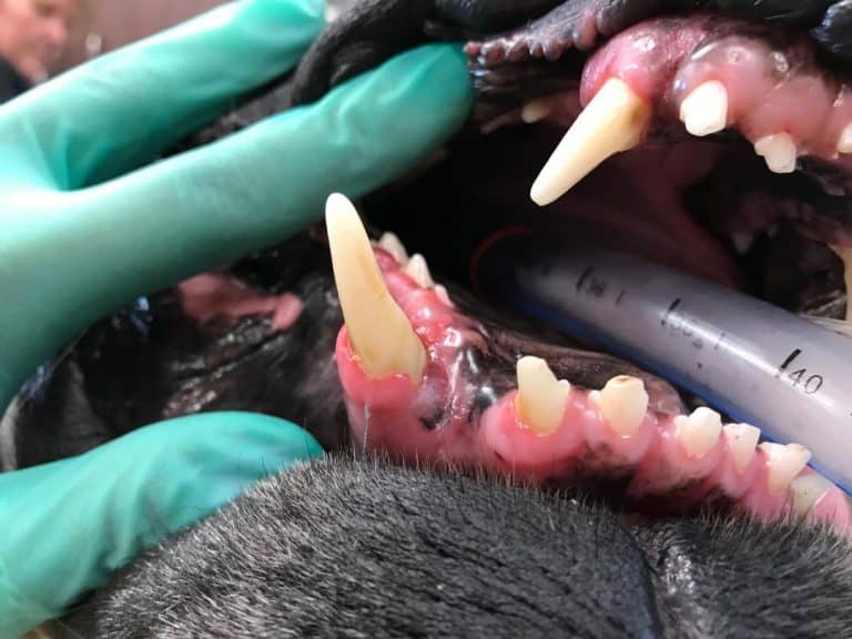
He presented with a painful luxated left upper canine tooth following trauma.

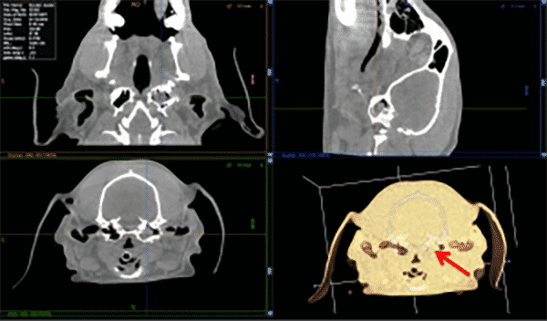
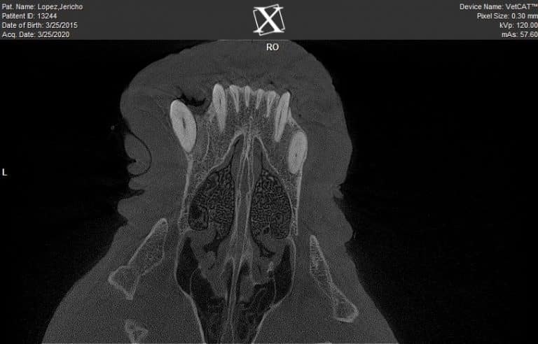
Pre-Op (above) VetCAT CT scan revealing extent of blockage in ear canal.

Sadly, Jericho’s tooth was displaced from its normal position in its socket for at least a couple of weeks- removing blood supply to the tooth at the time of his initial trauma allowing bone to begin to form in the socket. This can be easily seen on the VetCAT CT images taken pre-op.


Quite often a tooth like this can be placed back into its socket and secured with a splint and wire while the fractured bone heals, and a root canal procedure is performed at the time of splint removal to save the injured tooth. Since the tooth could not be reduced to its original place and it was periodontally unsound, it was surgically extracted.
While Jericho is missing a pretty large tooth, the VetCAT CT helped guide his vet specialist’s treatment decisions to quickly relieve Jericho’s painful injury.
Thanks to Dr. Patrick Vall, Veterinary Dental Specialist at Animal Dental Care & Oral Surgery in Colorado Springs, CO.
Download the case study PDF here to share: Jericho


