First seen in AuntMinnie.com
")
The Vital Role Radiologists Play in Facilitating Point-of-Care CT Imaging, a Retrospective Look
Today, point-of-care (POC) X-ray, ultrasound, and magnetic resonance imaging (MRI) imaging modalities are widely accepted by radiology departments. Such imaging at the patient’s POC—whether it be the intensive care unit (ICU), emergency department (ER), or the operating room (OR)—has become commonplace in hospitals across the United States and globally, as clinicians are able to obtain necessary diagnostic information quickly with minimal patient transport and discomfort.
Computed tomography (CT) imaging, on the other hand, could be seen as having had a slower acceptance rate of POC technology in the hospital. In the last few decades, advances in CT imaging have often meant that improved image quality comes with increasingly expensive machines. Nevertheless, radiologists in the U.S. and internationally have historically played a vitally important role in furthering access to POC imaging in the hospital, and they are poised to continue to do so in the future.
In partnership with radiology departments, Xoran Technologies understands the need for CT imaging directly at the point-of-care—it is generally more affordable, and if done right it does not require a dedicated room. Not only that, but POC CT also can mean a lower effective radiation dose for patients and a drastic reduction in transport risk for the critically ill patient or surgical candidate.
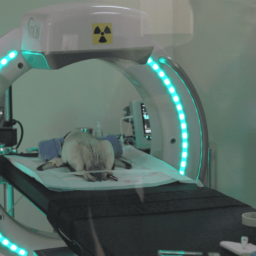
1996—A Landmark Use of POC CT Imaging
Some of the earliest documented cases of POC CT attempted to bring the CT imaging to the patient because of these very risks. Back in 1996, the University of Maryland Medical System in Baltimore, Maryland brought in a mobile CT scanner, the Tomoscan-M. It required two people to maneuver, but nevertheless, it was rated to bring excellent and good images about 92% of the time (1). The CT was used in the ICU and the OR, and in the discussion that followed, the radiologists noted:
Movement of patients receiving invasive monitoring and intensive care support, including the respiratory and circulatory systems, requires many specially trained personnel, such as respiratory therapists, nurses, and physicians, as well as considerable time to optimize safe transport for CT scanning when indicated. Requiring these personnel to leave the ICU floor decreases coverage for other ICU patients and is associated with significant cost per patient transport . Finally, the time required to transfer and scan critically ill patients may decrease the availability of CT for other patients, including those requiring acute CT scanning. (1)
The risks of intrahospital transport and the costs associated have been documented extensively since this 1997 publication in Emergency Radiology. Up to 80% of the time (2), an adverse event happens in which the patient has complications from the transport, including the adverse event of death. As these peer-reviewed articles show, there are additional workflow and economic benefits to the radiology department when CT is brought to the patient’s bedside.
2001—Identifying the Role of CBCT for POC Head Imaging
Since Xoran Technology’s beginning in 2001, the innovator and medical market leader in cone beam computed tomography (CBCT) imaging has been passionate about developing common sense, yet groundbreaking technologies that enable clinicians of all kinds to identify disease quickly and treat patients more efficiently and effectively.
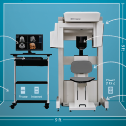
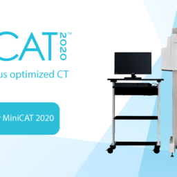
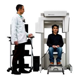
Xoran’s product line includes the upright MiniCAT™ in-office CT systems, the xCAT™ suite of truly mobile CT devices, a suite of veterinary 3D CT products, cloud-based image viewing through XoranConnect‘, as well as future grant-enabled projects in spine, lung, and AI.
After installing their first MiniCAT in 2004 in a California-based ear, nose, and throat clinic, Sacramento ENT, Xoran has become the market leader in POC CT imaging for ENT and allergy, with over 900 installations in the U.S. alone.
2008—The Evolution of the Imaging Industry
Xoran did not stop at innovating with our in-office upright CT. In 2008, Xoran introduced the truly mobile xCAT™ for ENTs to use intraoperatively. The Journal of Neuroradiology recognized the trend toward a proliferation in systems designed for head imaging and in 2009 published an article called “Review of Portable CT with Assessment of a Dedicated Head CT Scanner.” Looking at the CereTom, Tomoscan, xCAT ENT, and OTOscan, the authors noted the “obvious” risks of intrahospital transport and discussed both the workflow improvement and economic benefits to existing radiology departments when a mobile CT machine is implemented in the ICU and the OR settings. (3)
Fast-Forward to Today—POC CT Imaging at the Forefront of Radiology Innovation
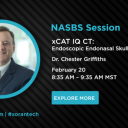
At the time in the late 2000s, the technology was such that imaging capability for brain imaging simply was not clinically sufficient. As components improved, Xoran was able recently to develop and release the xCAT IQ—”IQ” stands for image quality—this time with improved image quality with resolution detailed enough to visualize the brain.
The potential for improved patient care cannot be understated. Because the xCAT IQ is truly mobile, it easily maneuvers in small spaces and is uniquely suited for use in the ICU and intraoperatively in conjunction with surgical navigation and robotics, thereby helping to enable minimally invasive procedures and neuro and skull base surgical planning.
What’s more, because of the small footprint and fast scan times, the lower dose xCAT XL has a unique value proposition assisting neuro otologists in cochlear implant placement as well as in cranio-maxillofacial (CMF) procedures in which surgeons need intraoperative imaging to confirm fracture reduction and mesh placement.
Because Xoran’s CT images are DICOM 3.0 compatible, images can be sent quickly over the existing hospital PACS system, or using Xoran’s online image viewing and backup platform, XoranConnect‘.
Innovation Continues at Xoran. In 2019, Xoran announced it received a grant focused on integrated intraoperative imaging with surgical navigation from the National Institutes of Health (NIH) and the National Cancer Institute (NCI) through the Small Business Initiative Research (SBIR) project. The funds are facilitated by a matching grant from external investors, Decathlon Capital Partners.
This year, Xoran was awarded a grant from the National Heart, Lung, and Blood Institute (NHLBI) of the National Institutes of Health (NIH) to support the company’s research and development efforts for lung computed tomography (CT) imaging.
Leveraging its knowledge and experience in the field as the pioneer and medical market leader in cone beam CT, Xoran has proposed an open-bore, truly mobile CT to assist in the identification of lung disease as a part of the grant. In addition, Xoran has established collaborative partnerships with specialists in Pulmonary and Critical Care Medicine and Radiology at the University of Michigan, Ann Arbor.
Throughout these innovations, Xoran understands the importance of engaging closely with radiology departments to ensure that these point-of-care CT imaging systems are implemented in accordance with current radiology, medical physics, hospital administration, and IT guidelines. Radiologists will always be needed to ensure the safe, low dose, patient-centric implementation of radiologic procedures, and the interpretation of image results. Contact Xoran today at info@xorantech.com to continue the conversation.